Examples Of Skin Cancer Lesions / Skin Cancer Photos What Skin Cancer Precancerous Lesions Look Like - Melanoma contributes to 75% of deaths from skin cancer.
Examples Of Skin Cancer Lesions / Skin Cancer Photos What Skin Cancer Precancerous Lesions Look Like - Melanoma contributes to 75% of deaths from skin cancer.. 5 depending on location and size, the incision may be left to close on its own. 5% of total skin cancers diagnosed in the united states but causes most skin cancer deaths. Treatment may include corticosteroids, antibiotics, antifungal aids and other medications given systemically or topically. skin lesions may result from a wide range of causes, as harmless as, a small scrape or serious skin cancer. skin lesions can include, warts, skin tags, moles and skin cancers.
These conditions include moles, freckles, skin tags, benign lentigines, and seborrheic keratoses. _____ is the most dangerous form of skin cancer and is often characterized by black or dark brown patches and may appear uneven in texture, jagged, or raised. These types of cysts, lumps and bumps are common, which is why it does not pay to panic when you notice a bump on your dog. An image of a skin lesion (for example, melanoma) is sequentially warped into a probability distribution over clinical classes of skin disease using google. (a) dysplastic nevus, (b) seborrheic keratosis, (c) melanoma, and (d) squamous cell carcinoma (images publicly available in ).
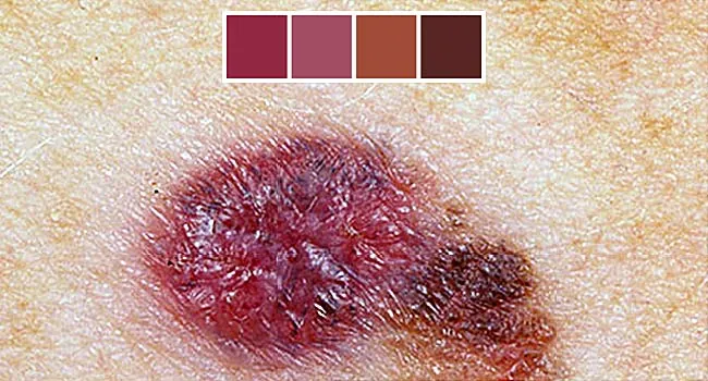
Melanoma Warning Signs And Images The Skin Cancer Foundation from 4ag46i294nta1038p13v77x1-wpengine.netdna-ssl.com (a) dysplastic nevus, (b) seborrheic keratosis, (c) melanoma, and (d) squamous cell carcinoma (images publicly available in ). 5 depending on location and size, the incision may be left to close on its own. A melanoma, we have to find or create a dataset that contains many examples of the things we want to detect. Tumor regression in pigmented lesions is a most interesting phenomenon occurring in both benign and malignant lesions. The deadliest form of skin cancer, melanoma (like the one shown above) is characterized by dark pigmentation, often with different shades of color, and irregular, asymmetric shapes. Since we want to design an algorithm that can identify skin lesions, e.g. Sores that do not heal; Changes which occur as a result of the natural development of, or due to external manipulation of the primary lesion.
This is the most common form of a premalignant skin lesion.
Melanoma skin cancer melanoma is a cancerous growth of melanocytes and most frequently develops in the skin.1 melanoma may also develop in other parts of the body that contain melanocytes including the meninges, the digestive tract, the eyes and lymph nodes.the following descriptions are limited to melanoma of the skin. Melanomas occur mainly on the skin but also on the mucosa of the oral, genital, and rectal regions and conjunctiva. It is caused mainly by excessive exposure to uv light, which damages dna. The pigmented lesion seen in the images is larger than most benign moles and also shows an irregular shape and multiple colors. Melanoma is considered the most dangerous form of skin cancer as it typically will spread to other areas of the body, including organs, if left untreated. Many people refer to a mole as any dark spot or irregularity in the skin, such as: Sores that do not heal; A reddish patch or irritated area, on the face, chest, shoulder, arm or leg that may crust, itch, hurt or cause no discomfort. With 5.4 million cases of skin cancer diagnosed each year in the united states alone, the need for quick and effective clinical screenings is rising 1, 2. Moles that change size, color or shape; These are non fatal, easily diagnosed and curable forms of skin tumors. These types of cysts, lumps and bumps are common, which is why it does not pay to panic when you notice a bump on your dog. 5 depending on location and size, the incision may be left to close on its own.
Therefore, due to the nature of skin cancer that should be diagnosed only with the. With 5.4 million cases of skin cancer diagnosed each year in the united states alone, the need for quick and effective clinical screenings is rising 1, 2. Follow the abcd's of skin cancer, they are: In fact, in up to 10% of cases, this condition can progress into a form of cancer called squamous cell carcinoma (scc). The deadliest form of skin cancer, melanoma (like the one shown above) is characterized by dark pigmentation, often with different shades of color, and irregular, asymmetric shapes.
Examples Of Suspicious Skin Lesions The Fi Rst Three Lesions In The Download Scientific Diagram from www.researchgate.net skin cancer is a disease in which skin cells grow out of control. (a) dysplastic nevus, (b) seborrheic keratosis, (c) melanoma, and (d) squamous cell carcinoma (images publicly available in ). There are different reasons why you may need lesion removal. Treatment may include corticosteroids, antibiotics, antifungal aids and other medications given systemically or topically. Basal cell carcinoma, squamous cell carcinoma, and melanoma. So it's important to check your skin once a month or so. Some of the common examples of secondary skin lesions are ulcers, erosions, scars, fissure, and crust. This is the most common form of a premalignant skin lesion.
Melanoma skin cancer melanoma is a cancerous growth of melanocytes and most frequently develops in the skin.1 melanoma may also develop in other parts of the body that contain melanocytes including the meninges, the digestive tract, the eyes and lymph nodes.the following descriptions are limited to melanoma of the skin.
Learn about the abcde assessment to detect melanoma skin cancer! Melanoma was the 19th most common cancer worldwide in 2008, with an approximate estimation of 200,000 new cases, and with the highest incidence rate in australia/new zealand, northern. Basal cell carcinoma, squamous cell carcinoma, and melanoma. Remember, melanoma can be cured if detected early, unlike many cancers. Tinea unguium is also known as? Melanoma accounts for only 4% of all skin cancers; Therefore, due to the nature of skin cancer that should be diagnosed only with the. It develops when you irritate your primary skin lesions by scratching. Malignant (cancerous) lesions or neoplasms. The previous two chapters dealt with bcc and scc — the two commonest types of skin cancer, both of which are keratinocyte derived. A melanoma, we have to find or create a dataset that contains many examples of the things we want to detect. Four examples of skin lesions: A pale patch of skin that grows.
Because the lesions of common benign disorders (ie, nevus, lentigo, acne, and seborrheic keratosis) and lesions of normal skin structures (ie, nose, ear) were observed in the primary training data set, we cropped and annotated these lesions based on image findings to create a secondary training data set (efigure 1 in the supplement). There are three common types of skin cancer: Melanoma is a type of skin cancer usually caused by excessive sun exposure, especially a history of sunburn (s). The deadliest form of skin cancer, melanoma (like the one shown above) is characterized by dark pigmentation, often with different shades of color, and irregular, asymmetric shapes. When the skin is exposed to the sun's ultraviolet radiation, lesions can develop on the skin.
Skin Cancer Photos What Skin Cancer Precancerous Lesions Look Like from img.webmd.com It may cause bleeding and crusting. This type of skin cancer is typically localized to the skin, and rarely spreads to other tissues. (sometimes the secondary changes make it impossible to see and describe the primary lesion) (scale, lichenification, keloid, excoriation, fissure, erosion, ulcer, atrophy, crust, hyperkeratosis) Treatment of skin lesions includes identifying the type of lesion (primary or secondary), the underlying cause of the lesion and the patient's health status. Tumor regression in pigmented lesions is a most interesting phenomenon occurring in both benign and malignant lesions. Suggested that the positive predictive value according to each type of skin lesions was not high, shown to be 72% for basal cell carcinoma, 49.9% for squamous cell carcinoma, and 33% for cutaneous melanoma. Below we will discuss a few groups of commonly acquired lesions. While lesions can occur on any site inside our bodies, one of the more visible places to incur lesions is on our skin.
The deadliest form of skin cancer, melanoma (like the one shown above) is characterized by dark pigmentation, often with different shades of color, and irregular, asymmetric shapes.
Benign lesions can be elevated (eg, a dermal naevus, dermatofibroma, or cyst), but a new elevated or thickened lesion may be suspicious for nodular melanoma or another form of skin cancer. Again, detecting and treating melanoma early is key to a good prognosis, as it can spread very quickly. Small skin cancers limited to the surface of the skin may not require treatment beyond an initial skin biopsy that removes the entire growth. (e.g., scalp, face and trunk are areas of high sebaceous gland concentration; When cancer is suspected, the lesion. By contrast, there are two lesions — actinic keratoses and intraepithelial carcinomas — that are keratinocyte precursors of scc. However, it causes the greatest number of deaths from skin cancer. If left untreated and allowed to spread more deeply into other tissues, melanoma can be fatal. However, moles are the most commonly examined for cancer if changes are detected. Diagnosing skin cancer begins with a visual examination. There are three common types of skin cancer: These are non fatal, easily diagnosed and curable forms of skin tumors. Four examples of skin lesions:
Generally, the greater the number of such lesions on the skin, the higher the risk of developing into cancer skin cancer examples. An urgent visit (within 2 weeks) to a see a dermatologist for closer examination with dermoscopy (a.


0 Comments